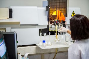
Ask the Expert: Battery Materials Analysis
During this Ask the Expert webinar, our team of battery materials experts will address your pre-submitted questions during the event, as well as present about our new battery lab’s capabilities.
Home » Complementary Techniques: X-Ray & SAM
X-ray imaging is a great non-destructive technique that is used in many industries to help visualize internal construction. Scanning acoustic microscopy (SAM) is another non-destructive technique that relies on ultrasonic sound waves to characterize internal construction, similar to how sonar and pediatric ultrasounds are performed. Both techniques rely on variations in material properties to generate contrast in the resulting images. This contrast helps distinguish different materials or identify structural anomalies.
As X-rays pass through materials, a certain amount is absorbed or scattered. The remaining X-rays that penetrate the material allow us to assess the material’s X-ray attenuation. By contrasting the number of X-rays that pass through with those that do not, a 2D image or 3D reconstruction can be generated.
As an ultrasonic sound wave travels through a stack of materials, a certain amount of reflection and transmission occurs at each interface the pulse encounters. The acoustic impedance of the two materials determines how much of the incident pulse is reflected at the interface and how much is transmitted into the next material. Reflections at different times can reveal thickness or material variations as the pulse propagates through the package. A 2D image can be created by analyzing reflected waves within a specific time range that corresponds to a particular layer in the device.
While both techniques are powerful non-destructive approaches that can help characterize internal features that aren’t optically visible, they are not interchangeable. Instead, they should be considered as complementary techniques where different information is portrayed in each image. X-ray can portray density and thickness variations that can help to characterize solder voiding. Scanning acoustic microscopy can show interfacial differences, down to submicron levels.
For example, if the solder bump was close to the expected thickness (and thus looked very good from a relative x-ray standpoint) but did not make contact with one of the pads, it would be classified as a false positive. Scanning acoustic microscopy, on the other hand, can detect submicron delamination within materials which makes it a great technique to identify open bumps, delamination and internal cracks.
Although the techniques are not interchangeable, there are situations when it is much more advantageous to use one over the other.
For example, in a dense area with extensive gold metallization, X-ray imaging may not provide much contrast. However, acoustic microscopy won’t be as adversely affected and thus may provide a more insightful image. Likewise, if there is an air cavity in a hermetically sealed package, sound waves would not be able to propagate past it, but X-rays would be unaffected.
Traditionally, combining the two techniques can provide more in-depth information than performing one or the other. For example, if there is a sheet of solder sandwiched by two metal plates, X-ray imaging could capture potential voids in the x-y plane. However, identifying where the voids are in the z plane will be challenging.
Reflective scans can give an idea of where the first void is along the propagation path, depending on the orientation of the scan. Most of the incident pulse is reflected at this point and thus there is no data collected beyond the void. By comparing the total voids (as seen in X-ray) and the first voids in the path (as seen in SAM), the location of the voids in the z direction can be inferred.
Both 2D X-ray imaging and acoustic microscopy are effective non-destructive testing methods.
They are based on different physical principles, offer varying imaging depths and resolutions, and are suited to different materials and applications. While SAM provides interfacial information, X-ray imaging offers an aggregate view. Ultimately, the choice between these techniques depends on the specific requirements of the imaging task, including the desired level of detail and resolution.
During this Ask the Expert webinar, our team of battery materials experts will address your pre-submitted questions during the event, as well as present about our new battery lab’s capabilities.
Biocompatibility assessments are a battery of tests performed to evaluate a product, medical device, or other material for the risk of biological hazards.
In this webinar we introduce Fractography which is a failure analysis evaluation technique when components fracture
May 15, 2024
Please join us for coffee and conversations! Enjoy a cup of coffee and pastries as you get to know our technical experts.
Home » Complementary Techniques: X-Ray & SAM
This section is the opening paragraph that shows how EAG relates to real life.
Text here
Text here
Text here
Automated Test Equipment (ATE) systems are carefully designed to test a wide range of semiconductor devices. EAG provides tailored ATE test solutions to meet the unique needs of clients.
In this webinar we introduce the application of Electron Microscopy to Lithium Ion Batteries from Micron to Atomic Level
Essentials of Particulate Testing: USP <788> and AAMI TIR42 While there are many standards available for particulate testing, USP <788> and AAMI TIR42 are the most important. This is especially
Demand for compound semiconductors has rapidly grown recently. During this live event we answered questions surrounding the Materials Characterization of GaN HEMT power transistors.
To enable certain features and improve your experience with us, this site stores cookies on your computer. Please click Continue to provide your authorization and permanently remove this message.
To find out more, please see our privacy policy.